D2001
2 year ago
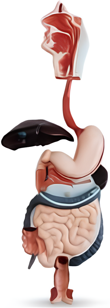
The Human Digestive System Model is an invaluable educational tool used to study and understand the complex processes of digestion in the human body. It provides a three-dimensional representation of the various organs and structures involved in the digestive system.
This model typically includes the following components:
Mouth: The model showcases the oral cavity, including the lips, teeth, tongue, and salivary glands. It highlights the initial stage of digestion, where food is chewed and mixed with saliva.
Esophagus: The esophagus, a muscular tube connecting the mouth to the stomach, is represented in the model. It demonstrates the peristaltic contractions that propel food from the mouth to the stomach.
Stomach: The model features a detailed depiction of the stomach, showing its shape, size, and position within the abdominal cavity. It may also illustrate the gastric glands responsible for producing stomach acid and enzymes.
Small Intestine: This section of the model represents the small intestine, which consists of three parts: the duodenum, jejunum, and ileum. It often highlights the villi and microvilli lining the walls of the small intestine, which increase the surface area for nutrient absorption.
In addition to the Human Digestive System Model, there are other medical training models available, including:
 Full Body Trauma Manikin
High Fidelity Simulation Model
Full Body CPR Training Manikin
Full Body Training Manikin
These models are designed to provide realistic training experiences for healthcare professionals and students in various medical scenarios. Whether it's practicing advanced maternity examinations, mastering surgical suturing techniques, or learning about the anatomy and physiology of different body systems, these models offer a hands-on approach to learning.
It's important to note that the use of these models greatly enhances medical education and improves patient care by allowing healthcare professionals to gain practical skills in a safe and controlled environment.
Features:
Model demonstrates alimentary canal from mouth to rectum in median section and display buccal cavity, pharynx, esophagus with half of the stomach, opened duodenum, small and large intestines, opened appendix, unfolded rectum, transverse colon, liver, pancreas, etc.
Similar Video Recommendation
You May Also Like
If you are interested in the product, contact Bossgoovideo.com for more information
- *To:
- Yinchuan Erxin Technology Co., LTD
- *Message:
-
Submit
Main Product:
First Aid Skill Training Model,
Clinical Skill Training Model,
Nursing Skill Training Model ,
Human Anatomical Model,
Stomatology Model,
lmaging Medicine Model


